August 17, 2015 - As MRI is increasingly used to diagnose breast cancer, researchers designed a study1 to determine the diagnostic performance of combined dynamic contrast-enhanced magnetic resonance imaging (DCE-MRI) and diffusion-weighted imaging (DWI) for breast cancer detection.
In this retrospective study, using PUBMED, EMBASE, Web of Science, and Cochrane Library databases, researchers did statistical analysis of sensitivity and specificity, positive likelihood ratio (PLR), negative likelihood ratio (NLR), diagnostic odds ratio (DOR), and diagnostic accuracy using the summary receiver operating characteristic (SROC). All analyses were conducted using STATA (version 12.0), RevMan (version 5.2), and Meta-Disc 1.4 software programs.
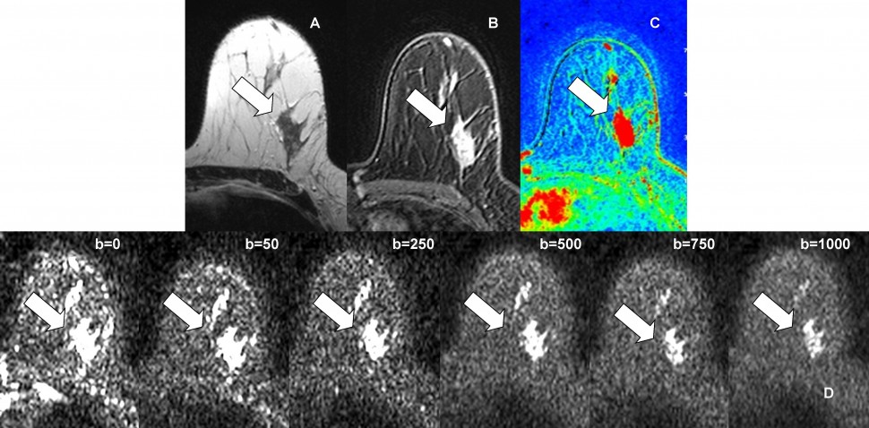
MRI images showing an IDC lesion in the left breast (axial plane). A – T2 sequence. B – T1 contrast enhanced image. C – Perfusion map (highly perfused regions appear in red). D – Different b-values diffusion-weighted images..2
They looked at a total of 1140 patients with 1276 breast lesions and found the pooled sensitivity and specificity of combined DCE-MRI and DWI were 91.6% and 85.5%, respectively. The pooled sensitivity and specificity of DWI-MRI were 86.0% and 75.6%, respectively. The pooled sensitivity and specificity of DCE-MRI were 93.2% and 71.1%. The area under the SROC curve (AUC-SROC) of combined DCE-MRI and DWI was 0.94, the DCE-MRI of 0.85.
Based on these results, the authors concluded that combined DCE-MRI and DWI had superior diagnostic accuracy than either DCE-MRI or DWI alone for the diagnosis of breast cancer.
Source:
- Zhang L, Tang M, Min Z, et al. Accuracy of combined dynamic contrast-enhanced magnetic resonance imaging and diffusion-weighted imaging for breast cancer detection: a meta-analysis. Radiology, Nuclear Medicine & Medical Imaging 72 out of 125.
- References: Department of Radiology, IPOLFG, Lisbon/ Portugal 2012


