February 8, 2016 - The ability of 18-fluorothymidine positron emission tomography/computed tomography (FLT-PET/CT) to identify primary tumors in gastric cancer is greater than that of contrast-enhanced computed tomography (CECT), and found FLT-PET/CT was better in evaluating regional nodal metastases, according to a recent study published in Abdominal Imaging.1
Since there has been no data available to date on the use of FLT-PET/CT for preoperative gastric cancer staging, and the authors designed a study to assess the value of FLT-PET/CT for preoperative gastric cancer staging in comparison with contrast-enhanced computed tomography CECT.
In a group of 96 gastric cancer patients, 96 FLT-PET/CT, 56 abdominal cavity CECT, and 51 resective operations were done. All three (FLT-PET/CT, CECT, and resective operation) were done in 29 patients. The results of FLT-PET/CT, CECT, and histopathological examinations were used to assess the ability of FLT-PET/CT and CECT to identify primary tumors, regional nodal metastases, and distant abdominal metastases. Assessment of regional lymph nodes was based on SUVmax in FLT-PET/CT and SAD (short-axis diameter) in CECT.
In the group of 56 patients examined with FLT-PET/CT and CECT, identification of the primary tumor was possible in 56 cases (100%) and in 53 cases (94.6%), respectively, (p = 0.013). Using ROC curve, the sensitivity and specificity of FLT-PET/CT in metastatic regional lymph node assessment were higher than those of CECT (p = 0.0033). FLT-PE/CT enabled identification of a greater number of extraregional abdominal metastases than CECT (n = 56; 19 vs. 15, respectively), but the difference was not statistically significant (p > 0.41).
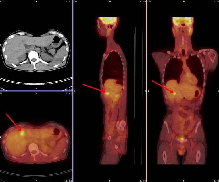
Image: 35-year-old male status post surgery, chemotherapy, and radiotherapy for rectal cancer. New lesion in the liver concerning for metastatic disease. PET/CT ordered for restaging.2
The authors concluded that FLT-PET/CT was more likely to identify primary tumors than that of CECT, and thus FLT-PET/CT was better in evaluating regional nodal metastases. Although, FLT-PET/CT enabled identification of a greater number of abdominal metastases than CECT, however, the difference was not statistically significant.
References:
- Staniuk T, Małkowski B, Śrutek E,,et al. Comparison of FLT-PET/CT and CECT in gastric cancer diagnosis. Abdominal Imaging. 02/01/2016.
-
Metastatic Colorectal Cancer. University of Virginia Health Sciences Center, Department of Radiology. http://www.med-ed.virginia.edu/courses/rad/petct/Colorectal.html.


