June 27, 2015 - Researchers found that 4D flow magnetic resonance imaging (MRI) flow-specific measures can be used to detect mild impairment of right ventricular (RV) dysfunction in primary left ventricular (LV) disease. However, this cannot be done using the conventional MRI and echocardiographic indices, according to a study1 from Journal of Magnetic Resonance Imaging.
Researchers acquired 4D flow and morphological 3T MRI data in 22 patients with mild ischemic heart disease who were stratified into two groups based on LV end-diastolic volume index (EDVI): lower-LVEDVI and higher-LVEDVI. They also performed 4D flow and morphological 3T MRI on 11 healthy controls. The RV volume was segmented at end-diastole (ED) and end-systole (ES). Pathlines were emitted from the ED volume and traced forwards and backwards in time to ES. The blood volume was separated into flow components. The Direct Flow (DF) component was defined as RV inflow passing directly to outflow. The kinetic energy (KE) of the DF component was calculated. Echocardiographic conventional RV indices were also assessed.
The results showed the RV 4D flow-specific measures “DF/EDV volume-ratio” and “DF/EDV KE-ratio at ED” were lower in the higher-LVEDVI group (38 ± 5%; 52 ± 6%) compared to the healthy (44 ± 6; 65 ± 7, P = 0.018 and P < 0.001) and lower-LVEDVI groups (44 ± 6; 64 ± 7, P = 0.011 and P < 0.001).
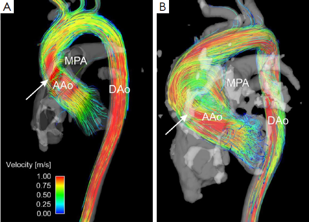
Image: 4D Flow Imaging with MRI.2
Based on the results, the authors concluded that primary LV disease mild impairment of RV function can be detected by 4D flow-specific measures, but not by the conventional MRI and echocardiographic indices.
References:
- Fredriksson AG, Svalbring E, Erksson J, et al. 4D flow MRI can detect subtle right ventricular dysfunction in primary left ventricular disease. J. Magn. Reson. Imaging 2015. DOI: 10.1002/jmri.25015
- 4D Flow Imaging with MRI. Cardiovascular Diagnostis and Therapy. Vol 4, No 2 (April 2014) /


