February 2, 2016 - Sonographic artifacts known as B-lines can been used to estimate alterations of lung parenchyma. According to a recent study,1 the number of B-lines on sonography were found to correlate with the extent of parenchymal changes on computed tomography (CT).
Multiple B-lines on sonography are seen in congestive heart disease, interstitial lung disease, respiratory infections, and neonates. The aim of this study was to compare the amount of B-lines on sonography to the extent of parenchymal changes on CT in children.
Lung sonography was performed on 60 patients aged 18 years and younger referred for chest CT at our institution. B-lines were counted from 5 anterolateral intercostal spaces bilaterally. The CT findings were documented and graded as absent, minimal, partial, or complete.
The number of B-lines on sonography increased consistently with the growing extent of parenchymal changes on CT. The differences in the B-line counts between the patients grouped according to the extent of parenchymal changes on CT were statistically significant except between patients with minimal and no changes (P < .01 Kruskal-Wallis and Tukey tests).
The authors found that the number of B-lines on sonography correlates with the extent of parenchymal changes on CT, and that various parenchymal changes were seen in patients with B-lines on sonography. B-lines were more frequently seen in patients with no changes on CT when imaged during general anesthesia.

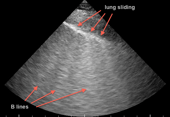
Image:
a. lung sliding – this indicates that there is no pneumothorax in the area you are scanning. b. there are B lines – this indicates that there is interstitial edema – this has no etiological information and must be coupled with the rest of the ultrasound and clinical examination to make a diagnosis. It could represent CHF, pneumonia, non-cardiogenic pulmonary edema, or any other interstitial process.2
References:
1. Martelius L, Heldt H, Lauerma K. B-Lines on Pediatric Lung Sonography: Comparison With Computed Tomography. JUM January 1, 2016 vol. 35 no. 1 153-157.
2. Thinking Critical Care. http://thinkingcriticalcare.com/tag/lung-ultrasound/


