July 30, 2015 – Researchers found ultrasound (US) is still effective when compared to positron emission tomography/computed tomography (PET/CT) during standard US surveillance of patients with breast cancer if internal mammary lymph node (LN) evaluation is routine, according to a recent study1 published in to the American Institute of Ultrasound in Medicine.
The authors wanted to compare [18F]fluorodeoxyglucose PET/CT to US based on pathologic results from US-guided biopsy and to evaluate the role of US in detecting internal mammary LN metastases in patients with breast cancer.
In the study, 37 patients with breast cancer underwent US-guided biopsy for suspected internal mammary LN metastases. Sensitivities for detecting internal mammary LN metastases were 76.7%, 96.7%, and 92.9% for initial US examinations, initial US combined with second-look US for initially missed cases, and PET/CT, respectively (P= .017). However, in a subgroup, there was a significant difference in sensitivities between initial and combined US (P = .019).
Based on the results, the authors concluded that while PET/CT is the best noninvasive method for evaluating internal mammary LN metastases, US is still useful if internal mammary lymph node (LN) evaluation is routine, and US-guided biopsies could be performed immediately on any suspected metastases and yield a high positive rate without serious complications.

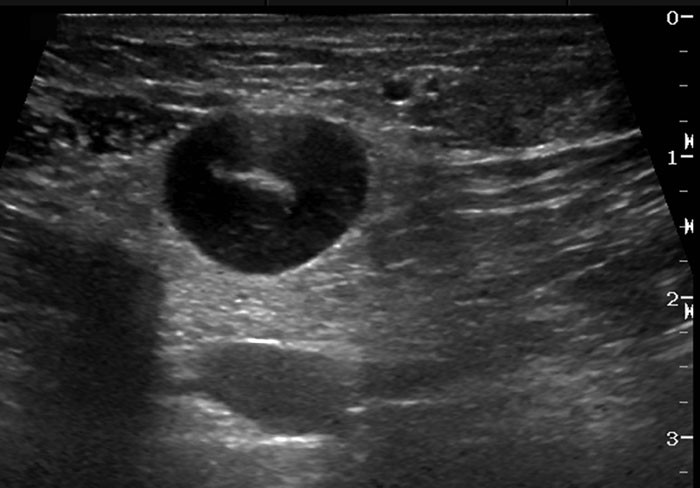
Image: Ultrasound of primary breast lymphoma.2
References:
- An YY, Kim SH, Kang BJ, Lee AH. Comparisons of Positron Emission Tomography/Computed Tomography and Ultrasound Imaging for Detection of Internal Mammary Lymph Node Metastases in Patients With Breast Cancer and Pathologic Correlation by Ultrasound-Guided Biopsy Procedures. doi: 10.7863/ultra.34.8.1385 JUM August 1, 2015 vol. 34 no. 8 1385-1394
- Radiology Case Reports. University of Washington in the public interest. ISSN 1930-0433. Vol 5, No 1 (2010). http://radiology.casereports.net/index.php/rcr/article/view/351/712.


