Jan. 2, 2015 - Magnetic resonance imaging (MRI) is a safe, reproducible imaging modality for human brown adipose tissue (BAT), according to a recent study published in January 2014 issue of the Journal of Clinical Endocrinology and Metabolism.
Manipulation of human brown adipose tissue (BAT) represents a novel therapeutic option for diabesity. The authors designed a study to use MRI to identify human BAT, delineate it from white adipose tissue, and validate it through immunohistochemistry. This technique could be used to help researchers develop therapies to battle obesity and related illnesses including diabetes.
A positron emission tomography and computed tomography (PET/CT) using (18)fluoro-2-deoxyglucose was scan performed post-surgically on a 25-year old female with hyperparathyroidism-jaw tumor syndrome. There was avid uptake within the patient’s mediastinum, neck, supraclavicular fossae, and axillae, consistent with BAT. Immunohistochemical staining using uncoupling protein-1 antibody was performed on one fat sample obtained from the suprasternal area during parathyroidectomy.

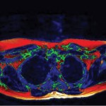
An axial MR image of the upper chest shows in green areas of potential brown fat in green. Image courtesy of the University Hospitals Coventry and Warwickshire and Terrance Jones, Ph.D.
Regions of interest (ROIs) identified retrospectively on MR corresponded to areas of high uptake on PET/CT. Eighty-three percent of the ROIs identified on PET/CT showed corresponding low MR signal. ROI were identified prospectively on MR based on signal intensity and appearance and compared with PET-CT. Eighty seven percent of 54 ROIs identified on MR showed a corresponding increased uptake on PET/CT. “The sample obtained at surgery corresponded with high uptake and low signal on subsequent PET and MR, respectively, and immunohistochemistry confirmed BAT,” the authors reported.
The authors concluded MR can be used to identify BAT in a living human adult with histological/immunohistochemical confirmation.
Reference:
Reddy NL1, Jones TA, Wayte SC, et al. Identification of brown adipose tissue using MR imaging in a human adult with histological and immunohistochemical confirmation. J Clin Endocrinol Metab. 2014 Jan;99(1):E117-21. doi: 10.1210/jc.2013-2036.


