March 15, 2016 – In patients with anaplastic gliomas, both 18F-fluoro-ethyl-tyrosine (18F-FET) and positron emission tomography (FET-PET) and magnetic resonance imaging (MRI) should be used preoperatively to assess a patients risks and estimate their prognosis, reports a recent study1 published in World Neurosurgy.
Although FET-PET imaging is an additional tool for tumor grading and surgery planning, not much is known about FET-PET imaging in anaplastic gliomas. The authors designed a study to assess the FET-uptake in anaplastic gliomas, compared to MR imaging, histopathological markers and its prognostic value.
Researchers retrospectively analyzed 46 patients (27m/19f) with an anaplastic glioma (WHO III) who received MRI and FET-PET imaging prior to surgery. Tumor volume was calculated in MRI and FET-PET imaging using a tumor-to-background ratio (TBR) and calculated maximum FET-uptake (TBRmax). They also calculated overall survival (OS) and histopathological markers (IDH1/2-mutation, oligodendrial differentiation, and Ki67 proliferation index). Univariate and multivariate analysis was performed for OS.
There was a significant correlation of TBRmax to OS and tumor volume in FET-PET imaging (TBR > 2.0) (P=.028) showed a higher correlation to OS than the volume of the contrast enhancing tumor part (P=.031). The highest correlation was observed for the intersection of volume TBR > 1.3 and the volume of the contrast enhancing tumor part (P=.005); FLAIR volume showed no significant correlation to OS (P=.401) in the univariate analysis. Anaplastic glioma with oligodendrial differentiation showed significantly higher TBRmax values (P=.029), while no significant difference was observed for IDH1/2-mutation (P=.752).
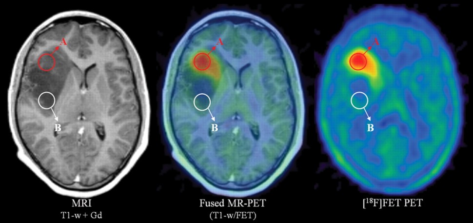
Figure. Combined [18F]FET PET MR clearly demarcate the anaplastic focus in a patient with heterogeneous glioma. Slightly increased contrast enhancement but highly increased tracer uptake proved this to be an anaplastic oligoastrocytoma WHO III (A), whereas nonenhancing tumor parts without significant tracer uptake were shown to constitute an astrocytoma WHO II (B).2
The authors concluded that static FET-PET provides significant prognostic information in anaplastic gliomas, which adds to the value of MR imaging, supporting the use of both modalities preoperatively to assess individual risks and estimate prognosis.
References:
1. Bette S1, Peschke P2, Kaesmacher J3, Delbridge C4, Pyka T5, Zimmer C3, Meyer B2, Ringel F2, Gempt J2. Static FET-PET and MR Imaging in Anaplastic Gliomas (WHO III). World Neurosurg. 2016 Mar 3. pii: S1878-8750(16)00342-9. doi: 10.1016/j.wneu.2016.02.094.
2. Molecular imaging of gliomas with PET: Opportunities and limitations. Neuro-Oncology. Oxford Journals. 2016. http://neuro-oncology.oxfordjournals.org/content/13/8/806/F3.expansion.html


