October 9, 2015 - A recent study found zoomed diffusion-weighted imaging (z-DWI) reduces image distortion and provides better registration accuracy with PET images than conventional echo planar imaging (EPI) DWI (c-DWI).
Researchers designed a study to compare zoomed diffusion-weighted imaging (z-DWI) with reduced field of view (FOV) by spatially selective radiofrequency pulses and conventional echo planar imaging (EPI) DWI (c-DWI) with regard to registration quality using positron emission tomography / magnetic resonance (PET/MR) in patients with malignant tumors.
Using a PET/MR system, the investigators conducted fludeoxyglucose (18F) PET imaging c-DWI, and z-DWI using simultaneously on 21 patients, with known or suspected malignancy on. They assessed registration accuracy between PET and DWI based on the area of maximum overlap and central point displacement of the tumor. EPI factor, echo time (TE), matching area, and displacement were compared between c-DWI and z-DWI by paired t-test. Agreement of apparent diffusion coefficient (ADC) acquired by the two sequences were also assessed with linear regression s and Bland–Altman plot analysis.
They detected thirty-two lesions on both PET and DWI. However, in all cases, EPI factor was smaller with z-DWI than c-DWI, registration accuracy was better with z-DWI in 30 of 32 lesions, and both average matching area and central point displacement were significantly improved.
The authors concluded that z-DWI reduces image distortion and provides better registration accuracy with PET images compared to c-DWI.

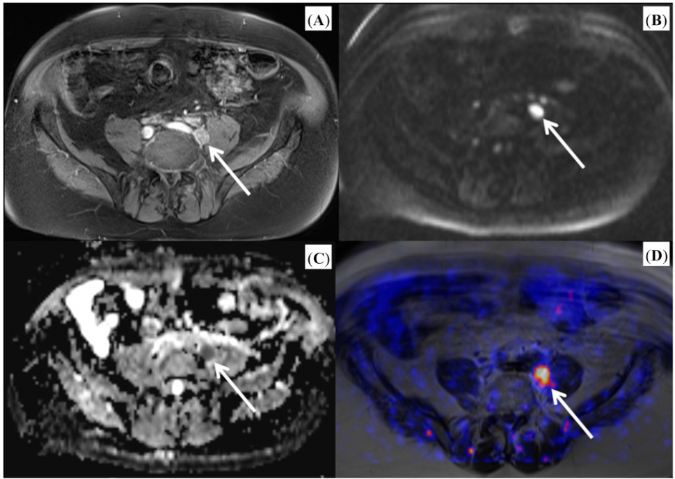
Image: Example of fused PET/MR image. This studies shows pronounced choline uptake of the lymph node. (Arrows indicate lymph node metastasis).2
References:
- Sagiyama K, Watanabe Y, Kamei R, etc. Comparison of positron emission tomography diffusion-weighted imaging (PET/DWI) registration quality in a PET/MR scanner: Zoomed DWI vs. Conventional DWI. J. Magn. Reson. Imaging 2015. DOI: 10.1002/jmri.25059.
- Molecular Research in Urology 2014: Update on PET/MR Imaging of the Prostate. Int. J. Mol. Sci. 2014, 15(8), 13401-13405; doi:10.3390/ijms150813401. http://www.mdpi.com/1422-0067/15/8/13401/htm.


