RSNA 2014 new technology review
By Cristen Bolan, Radiolopolis.com
Trends in radiology
Dec. 21, 2014 - Considered one of the top 10 medical advancements of the 20th Century, medical imaging already crossed new frontiers in science and medicine. Today, advances in computed tomography (CT), magnetic resonance (MR) and ultrasound imaging have substituted many interventional procedures—ultrasound has virtually replaced liver biopsies for patients with cirrhosis in Europe; CT has eliminated exploratory laparotomies because it is so accurate in identifying abdominal pathology; and MRI has made it possible to acquire a vast array of information about structure in the body.1
Recent advances in medical imaging have reached new frontiers in diagnosis and treatment., placing patients at the center of care. At RSNA 2014, from November 30 to December 5 in Chicago, we saw manufacturers providing solutions for some of the key clinical trends in radiology. These include breast tomosynthesis, CT lung screening, dose reduction, elastography, image-guided minimally invasive therapies, advances in neuroimaging with PET/MR technology, and point-of-care ultrasound.
Radiolopolis.com was on the exhibit hall floor talking to OEMs about how their innovations in technology are meeting these evolving clinical needs.
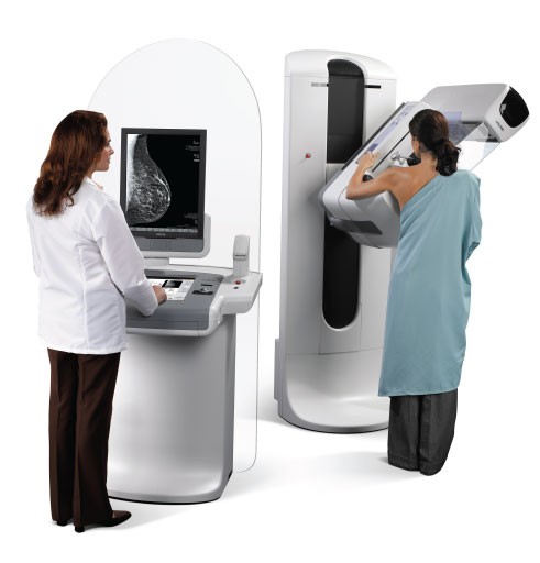
Breast Imaging
GE Healthcare – The SenoClaire and Invenia ABUS were the centerpiece of GE’s breast imaging solutions at RSNA this year.
The SenoClaire, a 3D breast tomosynthesis solution, helps clinicians find cancers in women with all types of breast tissue. With no increase in radiation dose compared to a standard 2D mammogram, SenoClaire uses a “step-and-shoot” method that reduces blur and increases image sharpness.
Invenia ABUS, automated breast ultrasound system, is the only technology approved by the FDA for screening the 40 percent of American women who have dense breast tissue. According to recent study,Assessing Improvement in Detection of Breast Cancer with Three-dimensional Automated Breast US in Women with Dense Breast Tissue: The SomoInsight Study, published in
Radiology, screening with ABUS in women with dense breasts improves breast cancer detection by an average of 27% over mammography alone.
For more information: www.gehealthcare.com or www.Reversitas.com
Hologic - Hologic introduced its 3D mammography exam as Genius 3D Mammography—the technology is designed to allow doctors to see masses and distortions associated with cancers more clearly than traditional 2D mammography. In a large peer-reviewed study published in the prestigious Journal of the American Medical Association (JAMA), researchers reported that Hologic’s 3D technology detects 41% more invasive breast cancers while significantly reducing anxiety-provoking callbacks due to false alarms. Hologic’s 3D exam is approved by the FDA as clinically superior to traditional mammography. The 3D mammography exam is available on the Selenia Dimensions platform.
Hologic also showed its C-View software option, which generates 2D images from the 3D data set. The majority of new Selenia Dimensions 3D systems ship with the C-View option. Also on display was the CE-marked and FDA-approved I-View software, an upgrade to any Selenia Dimensions system, I-View may combine 3D mammography and contrast enhanced 2D images to allow functional analysis.
For more information: www.hologic.com or www.Reversitas.com
Philips Healthcare - MicroDose SIacquires high image quality and low dose mammograms. MicroDose SI is based on photon-counting technology, which allows for the same low dose as the previous MicroDose model with an increase of up to 11 percent in image quality. MicroDose SI provides 50μm resolution, even in dense breasts. The Spectral Breast Density Measurement application is designed to help efficiently and objectively measure breast density. The system is also designed to improve the patient experience by providing women with a less stressful mammography experience. The breast positioning is easier and women also benefit from a curved support that enhances comfort.
For more information: www.philips.com/healthcare or www.Reversitas.com
Siemens Healthcare - The MAMMOMAT Inspiration Prime Edition digital mammography system lowers patient radiation dose by up to 30 percent compared to its predecessor model, depending on the patient’s breast-tissue thickness. The system replaces the scatter radiation grid common to conventional mammography with Siemens’ Prime algorithm for progressive image reconstruction, enabling lower radiation doses that are sufficient to generate high-quality breast images. The Opcomp function applies compression only as long as the patient’s breast is soft and pliable, stopping at the point of optimal compression. The MoodLight LED panel can create a more relaxing environment.
For more information: www.siemens.com or www.Reversitas.com
Computed Tomography (CT)
GE Healthcare – The manufacturer has built onto its Revolution CT new technologies, Revolution GSI, Revolution HD and Revolution EVO, designed to improve image quality, patient experience, greater economic value and advanced applications. Focused on lowering radiation dose, Revolution features a Gemstone Clarity detector, a dedicated 70 kVp scan mode for pediatric use and its ASIR-V iterative reconstruction solution. ASiR-V allows for routine imaging at up to 82 percent less dose. Spatial resolution is 20 percent higher than previous systems, and shows details as small as 0.28 mm. Revolution EVO gives flexibility to expand into advanced applications like TAVI planning, high heart rate CCTA and patients with implants.
For more information: www.gehealthcare.com or www.Reversitas.com
Hitachi Medical Systems America – SCENARIA Advanced 128 now features improved iterative processing technology (Intelli IP Advanced), enhanced workflow and Hitachi’s latest dose reduction features. SCENARIA can provide 128 slices per rotation for improved multi-planar reconstruction (MPR) visualization. With a high density view rate of 2,880 data samples per detector element per second, the SCENARIA Advanced 128 enhanced data collection rate over the 360º rotation provides improved data density on the periphery of the field-of-view. Making the shortest 0.35 second scan time available for a wide range of abdominal and long coverage exams in addition to cardiac exams. This standard feature on the SCENARIA 128 Advanced also enables scanning at a higher pitch resulting in faster exam coverage at a lower dose.
For more information: www.hitachimed.com or www.Reversitas.com
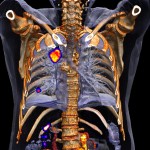
Siemens Healthcare – The new SOMATOM Definition Edge CT system is based on a TwinBeam Dual Energy X-ray tube enables simultaneous imaging at two different energy levels in a single-source computed tomography (CT). Dual-energy imaging technology provides tissue characterization applicable in clinical situations like the evaluation of kidney stones to differentiate between uric acid and non-uric acid stones, or when examining liver lesions, the material information contained in the dual-energy imaging acquisition can indicate liver lesions, with information that is based on the determination of contrast uptake in the tumor (Iodine Maps). The system also uses the new iMAR solution, a new iterative algorithm for metal artifact reduction.
A follow up study after a cardiac pump implantation – two volume rendering technique (VRT) images show that the severe artifacts caused by the implanted pump (left) are significantly reduced by iMAR reconstruction technique and the surrounding anatomical structures can be clearly visualized (right). Image courtesy of University Erlangen-Nuremberg, Erlangen, Germany.
For more information: www.siemens.com or www.Reversitas.com
Siemens Healthcare – The SOMATOM Force dual-source CT system delivers previously unachievable low-dose images with the benefit of ADMIRE (Advanced Modeled Iterative Reconstruction), the company’s newest iterative reconstruction algorithm. Used in conjunction with SAFIRE (Sinogram Affirmed Iterative Reconstruction), Siemens’ raw-data based iterative reconstruction algorithm, users can achieve a radiation dose reduction of up to 60 percent. ADMIRE uses additional modeling loops that help deliver higher resolution at organ borders and improve delineation of edges. With the combination of SAFIRE, ADMIRE and the technology in the SOMATOM Force, Siemens can deliver studies at very low doses that maintain high image quality and preserve a natural image impression.
For more information: www.siemens.com or www.Reversitas.com
Digital radiography (X-ray)
Canon - For medical facilities transitioning from CR to DR, Canon U.S.A., Inc., provides an economical approach with limited downtime with the availability of the RadPRO DELINIA 200 Digital X-ray Acquisition Cart. The cart transports the Canon CXDI-701C/801C/401C Wireless detectors with Auto Detection mode to provide health care organizations a cost-effective way to transition to DR technology. More information: www.usa.canon.com/dr or www.Reversitas.com
Canon - The RadPRO DELINIA 200 Digital X-ray Acquisition Cart comes equipped with a computer, access point, touch screen monitor, detector holder, and a choice of the Canon CXDI-701C, CXDI-801C, or CXDI-401C Wireless DR system. In X-ray Auto Detection mode, the detector will detect X-rays at exposure and shift to image acquisition mode automatically, without the use of a typical X-ray generator interface. As a result, the Canon wireless detector can deliver high-quality imaging and help accelerate exams by providing results within seconds, using the installed X-ray generator in an existing radiography room or a mobile generator, without the need for cabling or special interfacing.
For more information: www.usa.canon.com/dr or www.Reversitas.com
Canon - The new RadPRO OMNERA 400 Series of Digital Radiographic Systems by Canon U.S.A., Inc., has been designed for demanding hospital imaging departments to help create an efficient workflow to increase patient throughput. The RadPRO OMNERA 400A Auto-Positioning Digital Radiographic System incorporates fully automatic motorized positioning and features automatic stitching, a tube-mounted 10-inch touch screen, a vertical wall-stand and a table with extended range of motion. It will be outfitted with certain Canon CXDI Digital Radiography detectors, which are sold separately. Constructed of rugged crafted aircraft aluminum, the RadPRO OMNERA 400A System also provides the flexibility and ease of use helpful to healthcare professionals for delivering patient-focused care.
More information: www.usa.canon.com/dr or www.Reversitas.com
Fujifilm Medical U.S.A. – The lighter and more durable digital radiography detector the better. Fujifilm redesigned its FDR-DEVO II detectors, available in 24x30cm* Csl, 14”x17” and 17”x17”, to resist moisture and germs, are lightweight, and be more durable. LED status indicators and digital readout for connectivity, battery, impact sensing, and memory storage offer improved battery life compared to previous models. The detectors improved dose efficiency with DQE performance 20% with a 0.03 mR dose compared to prior models. Redesigned docking stand and two-slot chargers speed charging and display remote ready light.
More information: www.fujimed.comor www.Reversitas.com
GE Healthcare – The Optima* XR646, GE’s latest digital radiography system, handles larger patients with a rugged table that can lift patients up to 705 pounds with unlimited, or off-access, capacity. It moves in eight different directions and drops as low as 50 centimeters off the floor for easy access. Advanced features such as auto-tracking capability and auto image paste at the wallstand help increase productivity. The new touch-screen monitor produces images in less than three seconds allowing technologists to deliver fast results.
For more information: www.gehealthcare.com or www.Reversitas.com
GE Healthcare – The Discovery* IGS 740 is a rail-free, laser-guided premium interventional X-ray system for interventional radiology procedures. Its mobile gantry platform provides outstanding imaging flexibility while keeping the ceiling unobstructed to freely position the monitors, radiation shields and lights during interventions. The GE 41x41-cm detector (16.1 inches) offers a large field of view for imaging large anatomies at low dose. The wide bore C-arm makes it easier for clinicians to image the anatomy of interest and accommodate large patients. The dedicated arm-imaging positions facilitate arm fistulograms for hemodialysis patients, enabling full patient access from the left or right. For interventional oncology, clinicians can perform off-centered liver 3D acquisitions to support chemoembolization or Y90 procedures.
For more information: www.gehealthcare.com or www.Reversitas.com
Philips Healthcare - AlluraClarity Interventional Suites are tailored to neurology, oncology, and the hybrid OR.
- NeuroSuite deploys a combination of 20-inch and 15-inch flat detectors along with the latest 3D tools for effective device guidance. Tuck the small detector past the shoulder right up next to the head for a very short SID, enhanced image quality, and low dose.
- OncoSuite features the latest 3D image support for transarterial, such as TACE, and percutaneous interventions to enable the full range of IO care.
- Hybrid Suite – FlexMove provides a ceiling mounted C-arm that can be quickly moved anywhere it is required around the table, or completely out of the way if necessary. Suite deployment also includes either a Philips Xper Table or the Maquet Magnus table system.
For more information: www.philips.com/healthcare or www.Reversitas.com
Siemens Healthcare – Siemens’ new syngo Dyna4D software enables time-resolved 3D imaging in angiography, permitting 3D visualization of blood vessel volume as well as blood flow. The new syngo DynaCT SMART algorithm allows the clinician to remove metal artifacts from patient medical images, potentially enabling the detection of complications, such as hemorrhaging that occur in the vicinity of metallic objects contained within the patient’s body.
For more information: www.siemens.com or www.Reversitas.com
IT Solutions
Carestream – To manage the rincreased volume of datasets involved in reading breast tomosynthesis, new features on PACS designed to streamline reading DBT are critical to productivity. Carestream introduced its digital breast tomosynthesis (DBT) module digital breast tomosynthesis (DBT) module including a slabbing tool, improved workflow capabilities and the display of DICOM-compliant 2D synthetic views (which are generated from the 3D dataset).
The new slabbing tool combines slices of a DBT series, while allowing the user to choose different rendition modes and slab thicknesses. In scientific studies, radiologists have reported that this capability can help visualize calcifications and decrease reading time. The generation of 2D synthetic views is an alternate approach to acquiring conventional 2D mammography views, which can help reduce the radiation dosage a patient is exposed to while allowing full advantage of the benefits of digital breast tomosynthesis.
More information: www.carestream.com or www.Reversitas.com
Carestream – Clinical collaboration tools lets health care providers share data and work together in real time; break down walls between ancillary departments, sites and networks; and provide physicians with a single view of critical patient records and information. Carestream’s Collaboration Platform enables users to capture, archive, manage and distribute clinical data, such as images, videos, photos and reports related to the patient from different ancillary departments such as endoscopy and dermatology. The platform’s patient-centric structure matches the organization of electronic health records. This Web-enabled platform can be used for telemedicine and to provide specialty care where it’s traditionally not available. In addition, the platform also offers a secure digital patient portal that allows patients to download, view, store and share their medical imaging studies with physicians and specialists.
More information: www.carestream.com or www.Reversitas.com
Carestream – The latest version of Carestream’s radiology information system allows users import a patient’s clinical history so referring physicians can gain a more holistic view of the patient when making diagnostic and treatment decisions. The company also showcased its newest cardiology image management platform that sends a text message or email notification to a physician when a patient has a critical condition and a dashboard that provides data to assist with diagnosis of the current exam.
More information: www.carestream.com or www.Reversitas.com
Fujifilm Medical U.S.A. – Radiologists are using 3D models to shape the future of patient-centered care. By leveraging image overlay tools available on Synapse 3D, radiologists can use volumetric imaging to generate 3D models and quantitative analysis of organs and other parts of the anatomy for surgical treatment planning. Fujifilm showcased the ability of Synapse 3D to create 3D models. Synapse 3D offers more than 30 clinical functions to choose from, with many functions utilizing Fujifilm’s Image Intelligence capability.
More information: www.fujimed.com, http://3dimaging.fujimed.com/ or www.Reversitas.com
GE Healthcare - The new Centricity Solutions for Enterprise Imaging delivers a common viewing, analytics and vendor-neutral archiving (VNA) experience across specialties. Designed to support multiple care-specific workflows and offer clinicians the ability to work independent of location, GE Healthcare can now provide healthcare organizations with the tools they need to build Built on a common technology platform and leveraging industry standards, Centricity Solutions for Enterprise Imaging offers a modular, yet integrated solutions approach to unify imaging data. It creates a collaborative care environment by connecting acute, ambulatory and multi-specialty imaging needs with care pathway-specific workflows to help enhance diagnostic efficiency.
For more information: www.gehealthcare.com or www.Reversitas.com
Philips Healthcare – New at Philips is the DoseWise Portal, a comprehensive radiation dose management software solution aimed at managing radiation exposure risk to patients and their caregivers. The portal includes ClarityIQ, IMR and DoseAware. DoseAware provides real-time feedback on scattered X-ray dose so you can change your behavior. Users receive weekly or monthly dose reports automatically via email with your X-ray dose per procedure so you can identify your individual exposure trends. Procedural data on X-ray dose are sent in DICOM RDSR format to PACS or RIS to simplify data analysis.
For more information: www.philips.com/healthcare or www.Reversitas.com
Philips Healthcare – With the adoption of computed tomography (CT) lung screening for lung cancer detection taking place in the U.S., Philips introduced a new lung screening solution designed to offer health care providers a faster and more definitive pathway to lung cancer detection and treatment. The total solution is comprised of products and services to manage a comprehensive computed tomography (CT) lung screening program which tracks and guides patients across the health continuum.
The solution includes consultative services and marketing support for lung cancer screening services to help health systems reach high-risk patients. Control center software tools allow providers to follow a set of customized clinical protocols and establish interfaces with other hospital systems (HIS, RIS, EMR and PACS). Customizable workflow tools also send timely and accurate notifications, and track participant history and touch points.
CT lung image review and reporting offers specialized radiology software tools, which facilitate review and reporting of serial CT lung exams to identify and easily follow areas of concern. An online education portal measures and reports radiologist performance against a wide range of clinical cases, as well as structured education courses and other reference materials.
For more information: www.philips.com/healthcare or www.Reversitas.com
Siemens Healthcare – The new “teamplay” cloud-based networkaims to connect healthcare experts and increasing the usability of the wealth of medical imaging data. The platform helps link hospitals and health care experts to provide them with the ability to exchange data and pool their knowledge. Within hospitals, “teamplay” makes it possible to evaluate the extensive amount of information generated by imaging devices – e.g. scanner capacity utilization, examination times or radiation doses – and to compare the numbers against in-house and third-party reference values. This means imaging devices can be analyzed in close to real time and their operation optimized based on the results, right down to individual device level.
For more information: www.siemens.com or www.Reversitas.com
Siemens Healthcare - The latest version of Siemens’ syngo.via 3D and advanced visualization software supports physicians in treatment decision-making, planning, and assessment based on meaningful information – specifically for the field of oncology. Supporting the entire cancer care continuum across various modalities and departments, syngo.via is well-positioned to facilitate prompt, sound decisions and cost-effective therapy.
For more information: www.siemens.com or www.Reversitas.com
Siemens Healthcare - The latest version of syngo.plaza is an agile picture archiving and communications system (PACS) that joins 2D and 3D reading. With new scalable options, syngo.plaza grows with the customer’s clinical needs, helping to improve outcomes and keep initial investment low while allowing for future expansion. syngo.plaza also provides a wide range of applications and tools that support fast, efficient workflow. The high-throughput reading minimizes unproductive loading time and allows users to begin reading immediately. With syngo.plaza, it is easy to manage IT and to respond flexibly to changing requirements, helping administrators save time, resources, and effort. Additionally, the Smart Data Conversion feature enables a smooth transition when upgrading from existing systems.
For more information: www.siemens.com or www.Reversitas.com
Laser Printers
Carestream – The new CARESTREAM DRYVIEW 6950 Laser Imagerdesigned to produce rapid output of high-resolution images for all imaging modalities - CT, MR, CR, DR and other modalities – and supports output of full-field digital mammography (FFDM) and CR mammography images. It can deliver a maximum film density of 4.0, which is preferred for mammography.
More information: www.carestream.com or www.Reversitas.com
Molecular Imaging
GE Healthcare – Recently 510(k) cleared, SIGNA PET/MR is a time-of-flight (TOF) capable, integrated, simultaneous PET/MR system. This system represents a new chapter in helping clinicians achieve improved scan efficiency that may lead to more effective treatment paths for clinicians to offer their patients, particularly for oncology, neurology and cardiology treatment. The system features GE’s new MRI-compatible silicon photomultiplier detector (SiPM) technology.
This new digital detector is characterized by its exceptional timing resolution speed of 400ps, enabling what GE Healthcare calls TURBO TOF reconstruction. TURBO TOF offers highr SNR imaging while improving PET attenuation compared to non-TOF reconstruction systems. The SIGNA PET/MR also offers sensitivity of 21 cps/kBq enabling the potential for additional dose reduction or, alternatively, faster PET scans for the same dose. The system can be fully upgradable from a Discovery* MR750w 3.0T.
For more information: www.gehealthcare.com or www.Reversitas.com
GE Healthcare - Discovery IQ PET/CT is designed to deliver high image quality and Q.SUV, a quantitative SUV measurement for diagnosis, disease staging, treatment planning and assessment. It has the high sensitivity - up to 22 cps/kBq - the largest axial field-of-view is up to 26 cm and high clinical noise-equivalent-count-rate. Discovery IQ can image with both half the PET dose and half the scan time3. Q.Clear technology is a critical component of Discovery IQ.
For more information: www.gehealthcare.com or www.Reversitas.com
GE Healthcare – Discovery NM/CT 670 Pro with Q.Metrix advanced CT technology nuclear medicine by integrating with GE’s Optima CT 540. The system combines a 50-slice equivalent CT speed for extended fast coverage of time-critical scans with advanced CT applications with dose management tools, such as OptiDose, DoseWatch and AsiR, and a streamlined workflow for challenging stand-alone CT procedures. The NM/CT 670 Pro features Q.Suite for SPECT/CT quantitative accuracy has Q.Metrix, an application designed to measure and report SPECT standard uptake values in the organ or lesion of interest. Q.Metrix uses Q.AC, an image reconstruction technique for very low dose SPECT attenuation correction.
For more information: www.gehealthcare.com or www.Reversitas.com
Philips Healthcare – Improved contrast resolution and better SUV quantitation are the key advances in the Vereos PET/CT system. Digital Photon Counting (DPC) technology, which converts scintillation light directly to a digital signal coupled with time-of-flight performance have improved volumetric resolution, sensitivity, and quantitative accuracy 2 times compared to analog systems.
For more information: www.philips.com/healthcare or www.Reversitas.com
Siemens Healthcare - The Biograph mCT Flow PET•CT system overcomes the limitations of conventional bed-based, stop-and-go PET/CT imaging with FlowMotion, designed to move the patient smoothly through the system’s gantry while continuously acquiring PET data. The Biograph mCT Flow with FlowMotion enables imaging protocols based on the individual organ of interest, leveraging the finest volumetric resolution.The combination of a 78-cm bore with five-minute scanning and a continuous sense of progress throughout the scan offer a potentially more comfortable exam experience for the patient.
Accurate staging of lung cancer requires early detection of small lesions. Biograph mCT Flow™ allows for routine use of HD•Chest motion management techniques that enable delineation, as well as quantification of small lesions. Data courtesy of University of Tennessee, Knoxville, TN, USA.
For more information: www.siemens.com or www.Reversitas.com
Siemens Healthare - Symbia Intevo’s is an xSPECT system, combining the high sensitivity of SPECT with the high specificity of CT. Completely integrating data from both modalities, Symbia Intevo generates high resolution and reproducible quantitativeimages for SPECT. Siemens’ xSPECT modality reconstructs both the SPECT and CT portions of the image using the high-definition CT frame-of-reference for precise, accurate alignment that results in more clinical detail, which supports physicians’ ability to more confidently distinguish between degenerative disease and cancer.Symbia Intevo™ enables higher image resolution and more precise lesion characterization than conventional SPECT/CT bone imaging. Data courtesy of John Hopkins University, Baltimore, MD, USA.
For more information: www.siemens.com or www.Reversitas.com
Magnetic Resonance Imaging (MRI)
GE Healthcare – The new 510(k) pending SIGNA* Pioneer 3.T MRI system with MAGiC imaging solution is designed to enable clinicians to scan one more patient, every hour, every day. Developed in collaboration with SyntheticMR AB, MAGiC is engineered to help clinicians capture six image contrasts in a single scan in one-third the time. With MAGiC, clinicians will have the ability to generate multiple image contrasts in a single MRI scan including T1, T2, STIR, T1 FLAIR, T2 FLAIR and PD weighted images of the brain in a single acquisition. The contrast of images can be changed even after completing the scan by simply moving the cursor on the MAGiC interface to change acquisition parameters such as TR, TE and TI.
For more information: www.gehealthcare.com or www.Reversitas.com
Hitachi Medical Systems America - Oasis 1.2T a high field strength open MR offers high-field imaging with patient-centric features that minimize obstacles and delays associated with scanning challenging patients. Oasis combines patient comfort, high-field strength, efficient workflow and is reported to have a 99 percent uptime guarantee.
For more information: www.hitachimed.com or www.Reversitas.com
Hitachi Medical Systems America - Echelon Oval is a 74-cm oval bore is the widest 1.5T MR system available. Enhanced patient accessibility combined with Hitachi’s Workflow Integrated Technology (WIT), advanced imaging capabilities, and UltraPlus Customer Support, makes Echelon Oval an ideal solution for improved workflow, greater diagnostic confidence, and increased cost-efficiencies.
For more information: www.hitachimed.com or www.Reversitas.com
Philips Healthcare – The new Ingenia 1.5T S with Ambient MR In-Bore offers a soothing audiovisual experience to relax patients while inside an MRI scanner. The new MR system is designed for “First Time Right” imaging and for faster workflow, while enhancing the patient’s experience during MRI examinations. The system has fat-free and motion-free imaging techniques, patient-centric workflow and a unique patient experience during the exam. AutoVoice allows technologists to provide clear instructions for patients, and scanner noise is reduced through ComforTone scan techniques.
Premium IQ provides more diagnostic information in the available timeslot. (PRNewsFoto/Royal Philips)
For more information: www.philips.com/healthcare or www.Reversitas.com
Siemens Healthcare – The company debuted the MAGNETOM Amira 1.5 Tesla MRI system. Designed with the same technologies that are available on Siemens’ flagship MRI systems, yet the operating costs of the MAGNETOM Amira are much lower thanks to the new “Eco-Power” technology, which enables significant power savings in standby mode. MAGNETOM Amira is intended to meet the requirements of radiology practices, small and medium-sized hospitals, and larger facilities that are interested in a scanner to complement their existing systems. To enable high diagnostic reliability and patient comfort, MAGNETOM Amira is equipped with Siemens’ latest applications and syngo MR E11 software architecture. The system’s software platform includes the “Quiet Suite” technology, which is designed to minimize system noise during an MRI examination.
For more information: www.siemens.com or www.Reversitas.com
Ultrasound
Carestream – Enhnaced image quality is the value proposition for new ultrasound technology and that was Carestream’s aim with its debut of the its first ultrasound system—CARESTREAM Touch Ultrasound System. The new ultrasound platform delivers an all-touch control panel, integrated GPU processing power and smart transducer technology coupled with a single-board system design. This creates a highly reliable product with advanced imaging capabilities, a compact footprint and a modern user interface.
The all-touch control panel blends is designed to combine the speed and flexibility of a soft-user interface with the tactile feedback of traditional keys. Etched marking for primary controls assists the user in locating key functions without looking away from the image display monitor; maneuverability and a small, lightweight footprint makes the imaging process faster and easier; a “Swipe and go” badge log-on saves time and promotes secure access; a “Smart connect” transducer technology enables easy one-touch selection of the desired transducer; and it has a cold boot time is 18 seconds.
More information: www.carestream.com or www.Reversitas.com
GE Healthcare – The introduced a new software enhancement for its LOGIQ E9 ultrasound system called Shear Wave elastography. The new expert tool is designed to generate a precise, quantifiable measure of tissue stiffness, providing clinicians with accurate and consistent data, which may be used to identify underlying disease. Stiff tissue found during routine physical exams can be an early indication of disease and a gradual change in liver tissue stiffness can indicate the progression of liver fibrosis. Shear Wave’s power to provide a quantifiable measure of tissue stiffness may help enable clinicians to assess disease in the liver, breast and other organs immediately.
For more information: www.gehealthcare.com or www.Reversitas.com
GE Healthcare – Venue 50 portable tablet-style portable ultrasound delivers crisp images quickly with the simplicity of a tablet. The intuitive touch user interface offers easy gel-and-go scanning – a clinician can just select the probe and preset in one step. Designed for speed, it takes just moments to boot up and has no buttons, keyboard or knobs to slow clinicians down when scanning the patient or disinfecting the system.
The Vscan with Dual Probe is a pocket-sized ultrasound with a 2-in-1 probe, allowing clinicians to see both shallow and deep views of the body without changing probes. This latest innovation enables efficient triage and fast workflow, which may lead to time and cost savings in point-of-care settings.
For more information: www.gehealthcare.com or www.Reversitas.com
Fujifilm Medical U.S.A. – Point-of-care ultrasound with high visualization are designed to meet the needs of busy radiology departments looking for ideal portable ultrasound solutions. Fujifilm introduced the X-Porte kiosk designed for optimizing user workflow and varied environments, with adjustable height and gesture-driven touchpad for use in a standing or sitting position. Behind X-Porte is a new imaging technology used in ultrasound systems: SonoSite's proprietary Extreme Definition Imaging (XDI) technology. XDI is a new beam-forming algorithm that significantly reduces the visual clutter from side-lobes that affect all ultrasound products regardless of system size. The resulting ultrasound image appears cleaner with optimal tissue differentiation. The technological innovations are a result of the feedback and experiences of hundreds of clinicians that were observed during the development of the ultrasound kiosk.
For more information: www.fujimed.comor www.Reversitas.com
Samsung – The PT60A ultrasound is designed for all point-of-care applications. Needle Mate delineates needle location when performing interventions such as nerve blocks, corticosteroid injections and PIC line insertions. Easy needle detection improves accuracy in biopsy procedures. SDMR technology aims to eliminate speckle noise, while providing high contrast resolution in combination with enhanced edge definition for unsurpassed image quality.
For more information: www.samsung.com/global./business/healthcareor www.Reversitas.com
Samsung – The new HM70A was also on display at RSNA. It combines 2D and color image quality with fast frame rate. Needle Mate delineates optimal needle location when physicians perform interventions with pinpoint precision. Equipped with 4D and 3D XI, HM70A can be used for both baby facing and diagnostic 3D/4D imaging. ElastoScan applies strain imaging. High definition volume imaging uses a filter that removes unwanted speckle and noise while improving the visualization of edges and small structures in 3D data.
For more information: www.samsung.com/global./business/healthcareor www.Reversitas.com
Samsung – Ultrasound is an important tool in women’s health, and Samsung introduced the WS80A with Elite ultrasound system. With upgraded 3D imaging engine, clearer and more detailed images, and advanced S-Vue transducer technology provides enhanced vision of the scanned areas. 5D NT confirms fetal mid-sagittal view automatically from volume data, and the fetal nuchal translucency thickness is measures with simple and efficient semi-automatic operation. 5D LB detects and measures fetal long bones from volume data. 5D CNS is fetal brain measurement offering 6 measurements. 5D Stereo Cine, life-like 3D images can be displayed on Samsung 3D Smart TVs.
More information: www.samsung.com/global./business/healthcareor www.Reversitas.com
Siemens Healthcare - The ACUSON X600 ultrasound system is the newest addition to Siemens’ line of mid-range ultrasound products. An affordable multi-purpose solution, the ACUSON X600 system comes equipped with state-of-the-art technologies and workflow efficiencies migrated from premium systems. Advanced imaging solutions, such as real-time spatial compounding and Dynamic TCE tissue contrast-enhancement technology, reduce image artifacts and enhance border detection, offering improved detail and contrast resolution. Knowledge-based productivity applications simplify exam workflow and reduce keystrokes, enabling consistent measurements in less time while decreasing operator fatigue and risk of repetitive strain injuries. Three new volume transducers provide enhanced 3D/4D image quality for clear visualization, especially in OB/GYN. Capable of being upgraded when needs and budgets change, the ACUSON X600 system will continually benefit from software and hardware technologies pioneered on premium systems.
For more information: www.siemens.com or www.Reversitas.com
Siemens Healthcare - The new 2.0 release of the ACUSON X700 ultrasound system has an advanced imaging engine that boost efficiency and workflow, in addition to providing rapid, uniform visualization. The 2.0 release of the ACUSON X700 system includes eSie Touch elasticity imaging for noninvasive relative tissue stiffness analysis. Enhanced transducer compatibility and customizable upgrades make updating easy as clinical needs evolve.
For more information: www.siemens.com or www.Reversitas.com
References: Nikki. Top Ten Medical Advancements of the 20th Century. Posted November 21, 2008.


